
What Is Normal Posture? The Science Behind Healthy Spinal Alignment
You have probably been told to “sit up straight” or “stop slouching” more times than you can count. But has anyone ever explained what normal posture actually is, and why it matters beyond looking confident? In this article I want to answer that question with real science behind it, because it directly shapes how we approach your care here.
A common saying when learning something new is that if everything is important, then nothing is. The same can be said about the word “normal.” If everything is normal, then nothing is. Just like everything in healthcare, in order to define something as abnormal, we must first have an established normal to compare it to. So let’s start there.
Establishing a Science of Normal Posture
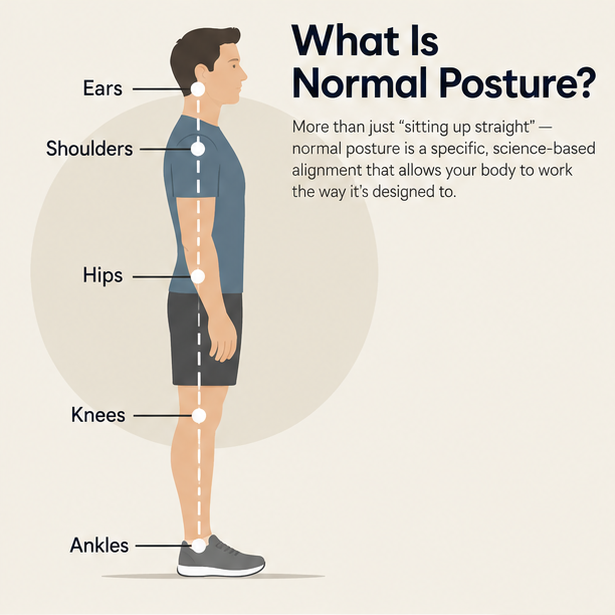
In our osteoporosis and posture article, I described a posture check where you use the position of the ears to shoulders, shoulders to hips, hips to knees, and knees to ankles to evaluate your posture from the side. When these five body landmarks line up vertically, one over the other, they describe the optimal arrangement of your body’s major weight-bearing masses with the line of gravity. This approach is often attributed to Kendall and colleagues in the book Muscles: Testing and Function with Posture and Pain.
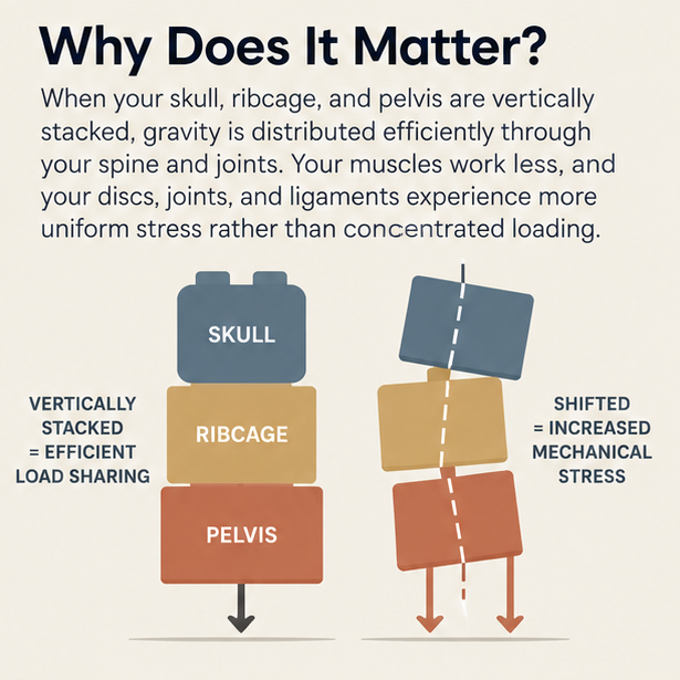
Your skull, ribcage, and pelvis are the three largest segments of your body. Each one has mass, and gravity acts on each of them constantly. When their centers of mass are vertically stacked, front to back and side to side, the compressive load of gravity passes through your spine and joints most efficiently. The muscles that hold you upright work less, and the discs, joints, and ligaments experience a more uniform distribution of stress rather than concentrated loading at specific points.
Biomechanics researchers White and Panjabi established this framework formally in the 1970s for the individual segments and joints of the spine, and Harrison later extended the concept to normal upright human posture in the 1980s. Harrison describes normal upright posture as zero rotations and zero translations of the skull, ribcage, and pelvis in any plane. Said differently, the skull, ribcage, and pelvis cannot be tilted or shifted away from neutral and still be considered normal. Think of a stack of Lego blocks with one piece shifted out of line. When any segment tilts or shifts away from the vertical stack, gravitational load is no longer distributed evenly, and the further a segment drifts from neutral, the greater the mechanical demand on every structure above and below it.
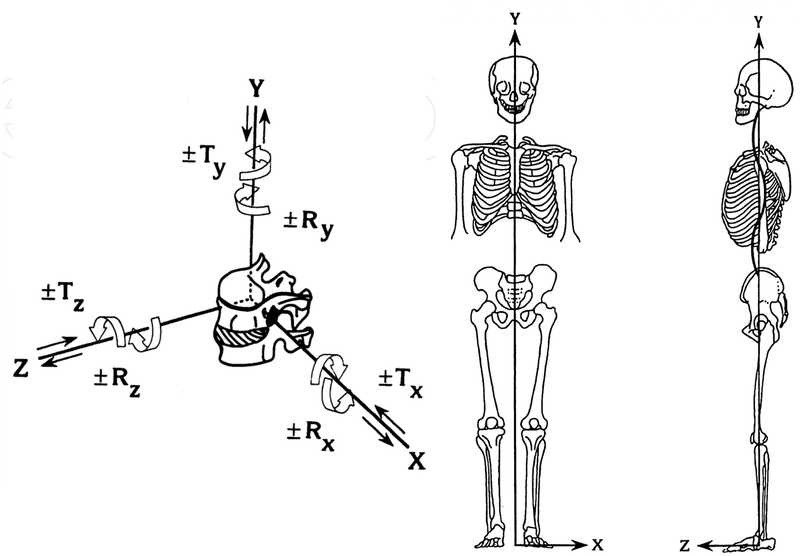
On the left: White and Panjabi’s model of segmental spinal alignment and movement (from Harrison & Oakley 2022)
On the right: Ideal structural / postural alignment as described by Harrison (from Harrison 1992 in Sweere: Chiropractic Family Practice)
In reality, nobody is perfect. Just as it is unreasonable to expect a blood pressure of exactly 120 over 80 at every moment, it is unreasonable to expect perfect posture at every moment. The key takeaway is that the further things shift away from neutral, the more stress builds at specific points, and that stress leads to secondary conditions such as chronic pain, loss of mobility, and degeneration over time.
Normal Posture Requires Normal Spinal Curves
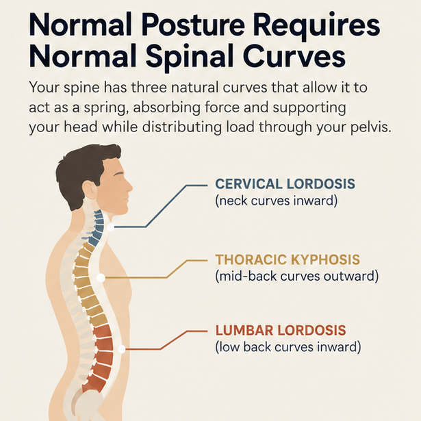
The vertical alignment described above is only possible because of the spine’s three natural curves. The neck curves inward (cervical lordosis), the mid-back curves outward (thoracic kyphosis), and the low back curves inward again (lumbar lordosis). These curves allow the spine to act as a spring rather than a rigid pole, absorbing force and supporting the weight of the skull above while the pelvis distributes load below.
The normal shape and depth of these curves has been studied extensively. The Harrison Spinal Model was developed through research on hundreds of asymptomatic subjects, meaning people with no pain or degeneration. It established that the cervical lordosis follows a circular arc geometry, a C-shape, while the thoracic kyphosis and lumbar lordosis follow similar mirroring elliptical arcs, J-shapes.
When a curve is lost, reversed, or excessive, the efficient load sharing the spine is designed for breaks down. A straightened or reversed neck curve, known as cervical kyphosis, will often shift the head forward relative to the shoulders. For every inch the head translates forward, the effective load on the cervical spine roughly doubles. Research by Breig showed that a forward head shift or straightened neck curve also places progressive mechanical tension on the spinal cord itself, much like stretching a rubber band until it pulls thinner. As we covered in our article on why your neck may be the reason your low back pain won’t go away, this consequence reaches well beyond neck pain and has even been implicated in radiating low back and leg pain.
Posture and Spinal Structure Are Related, But Not Quite the Same Thing
This is one of the most important distinctions I make during our initial and progress evaluations here at Structural Chiropractic.
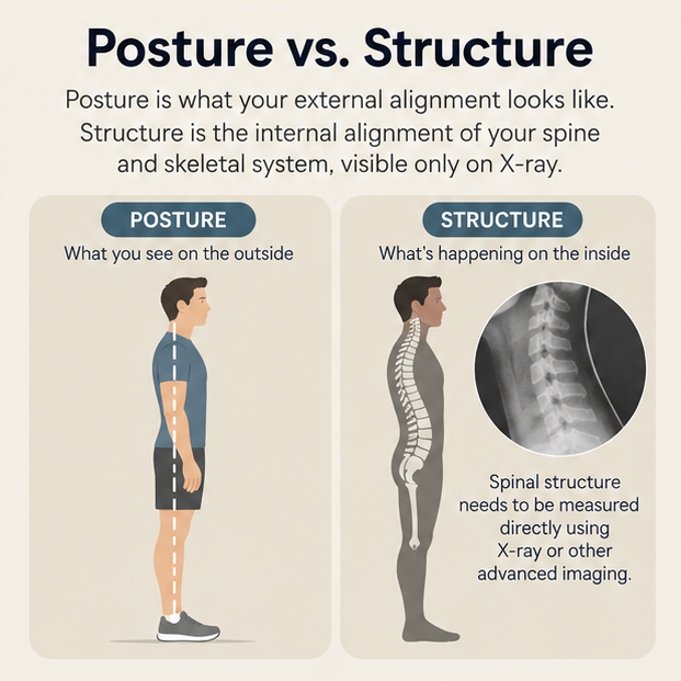
Posture refers to what your external body shape and alignment looks like while you are standing in space. Structure refers to the internal shape and alignment of the individual bones and joints of your spine and skeletal system. We can only see spinal structure on x-ray or an equivalent advanced imaging technique. The two are clearly related, since your spinal structure ultimately dictates the outward expression of your posture. However, it is impossible to predict your exact internal spinal structure from your external postural appearance alone.
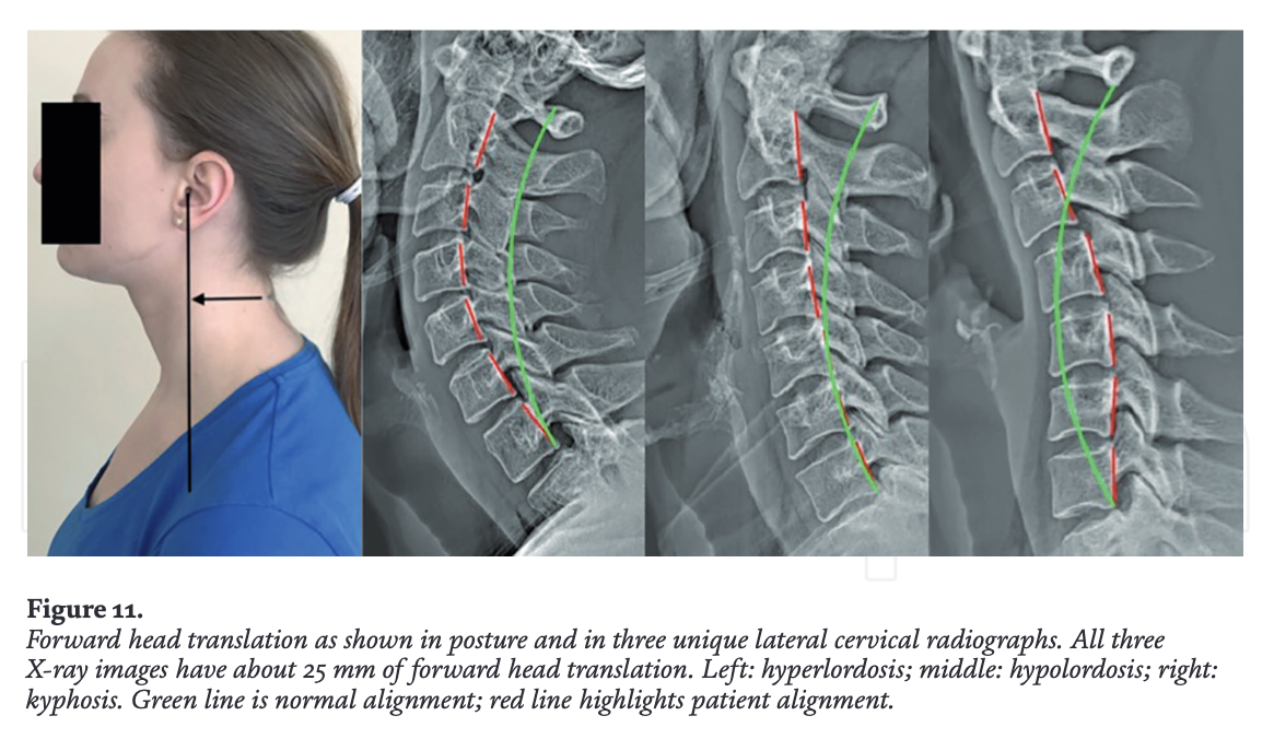
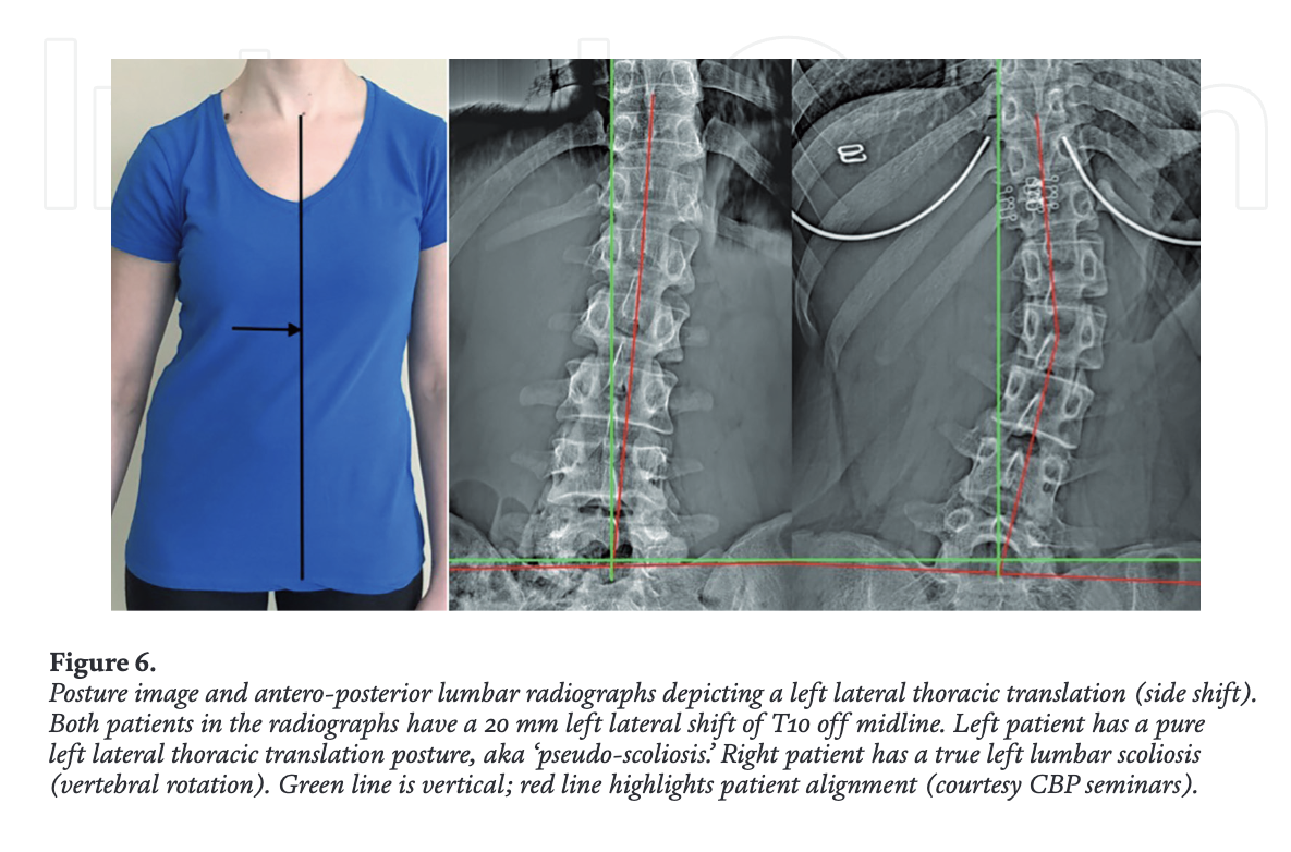
Someone can appear to stand reasonably straight while their cervical curve is reversed on x-ray. Two people can both show a lateral trunk lean, with one caused by a scoliosis and the other by a lateral thoracic translation, also called pseudoscoliosis. See Figures 11 & 6 below for 2 examples from Harrison & Oakley 2022). Posture gives us important information and can even be a tool to indirectly monitor structural progress during care. Ultimately, spinal structure needs to be measured directly with advanced analytical imaging in order to determine the exact quality and quantity of structural alignment or misalignment.
How We Objectively Measure Structure and Posture
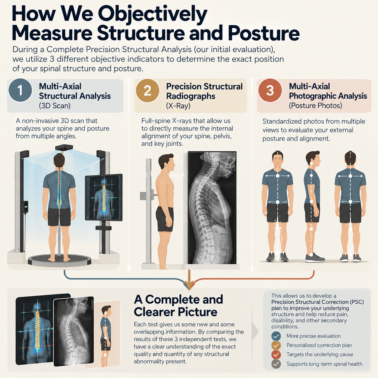
During a Complete Precision Structural Analysis, our initial evaluation, we use three different objective indicators to determine the exact position of your spinal structure and posture.
Multi-Axial Structural Analysis (3D Scan). A non-invasive 3D scan that analyzes your spine and posture from multiple angles.
Precision Structural Radiographs (X-Ray). Full-spine x-rays that allow us to directly measure the internal alignment of your spine, pelvis, and key joints.
Multi-Axial Photographic Analysis (Posture Photos). Standardized photos from multiple views that evaluate your external posture and alignment.
Each test gives us some new information and some overlapping information. By comparing the results of these three independent tests, we have a clear understanding of the exact quality and quantity of any structural abnormality present. At that point, we can develop a Precision Structural Correction (PSC) plan to improve the underlying structure responsible for posture, pain, disability, and a number of other secondary conditions.
If this article has you thinking about your own posture and spinal structure, a complimentary consultation is a great place to start. Call us at (412) 835-4844, visit structuralchiropit.com to learn more, or simply tap the button below to schedule your complimentary consultation.