
Why Can't I Take a Full Breath? Your Posture May Be the Reason
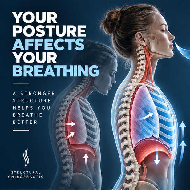
A patient came in recently whose primary concern wasn't neck pain or back pain. It was shortness of breath. Not an acute episode, but a problem they had noticed getting slowly worse over a number of years, primarily attributed to sitting at a desk and looking down at a phone or computer for hours every day. Over time, they noticed they couldn't take as full a breath as they used to, and it was affecting their energy and daily life.
As we talked through their history and reviewed their structural findings, one thing became clear: significant Anterior Head Syndrome, with the head shifted well forward of the shoulders and torso. When I explained the connection between head posture and breathing, I used a simple demonstration. I'll ask you to try it yourself right now.
Sit or stand tall, ears over shoulders. Take the deepest breath you can. Now shift your head as far forward as possible and try again. The difference should be immediate and unmistakable.
That difference has a physiological explanation, and it's backed by multiple published studies.
How Forward Head Posture Restricts Your Lungs
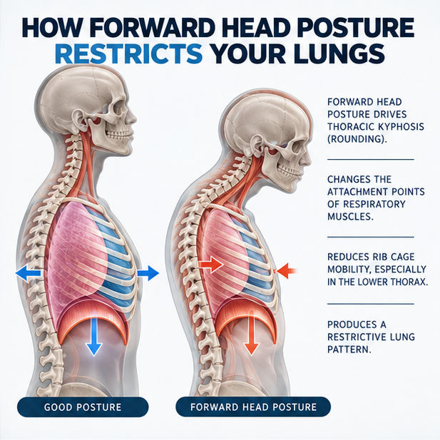
Your ability to breathe fully depends on your rib cage expanding freely in three dimensions with every breath: front, back, and sideways. That expansion is driven by the respiratory muscles, the diaphragm, the intercostals between the ribs, and accessory muscles in the neck and upper chest. Nearly all of these muscles have attachment points on the cervical and thoracic spine.
When the head shifts forward and the cervical curve is lost, every one of those attachment points changes. The muscles no longer function from an optimal position.
A biomechanical review published in the Indonesian Journal of Physical Medicine and Rehabilitation (Triangto et al., 2019) describes this mechanism precisely: Anterior Head Syndrome drives the thoracic spine into kyphosis, which reduces rib cage mobility and alters the attachments of the sternocleidomastoid, intercostal, and diaphragm muscles, producing what is clinically classified as a restrictive lung pattern.
The lower thorax is most affected. Research using 23 postural markers on the chest wall (Koseki et al., 2019) demonstrated that in the forward head position, the lower thorax shifts inward and its mobility during breathing is significantly reduced, even when the upper thorax is still moving. The lower lobes of the lungs, where most gas exchange occurs, are the ones most restricted.
What the Research Measured
These findings were measured using spirometry, the same standardized breathing tests used in clinical (like our practice) and research settings.
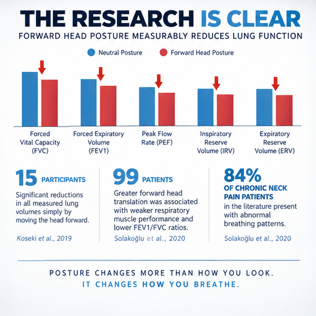
In a study of 15 participants who performed spirometry in both neutral and forward head positions (Koseki et al., 2019), forced vital capacity, forced expiratory volume, peak flow rate, and both inspiratory and expiratory reserve volumes were all significantly lower in the forward head position. These were instantaneous changes produced simply by shifting the head forward while seated.
A larger cross-sectional study of 99 chronic neck pain patients (Solakoğlu et al., 2020) confirmed this in a clinical population. The greater the forward head translation measured on cervical X-ray, the worse the respiratory muscle performance. Reduced FEV1/FVC ratios and forced expiratory flow rates followed the same pattern. Notably, 84% of chronic neck pain patients in the literature present with abnormal breathing patterns, suggesting this connection is the rule, not the exception.
Try It Right Now
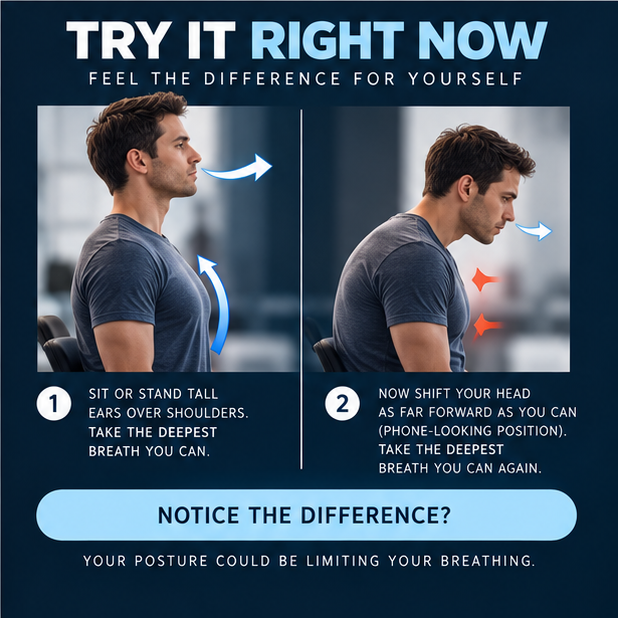
Do the test again with intention this time.
1. Sit or stand tall, ears over shoulders, chin gently back. Take the deepest breath you can.
2. Now shift your head as far forward as you can, the position most people hold when looking at a phone. Take the deepest breath you can again.
Notice the difference? That's your rib cage mechanics changing in real time. The lungs haven't changed. The airway hasn't changed. The structural position of the head and thorax has, and that's enough.
What This Means Clinically
If you or someone you know has been dealing with shortness of breath, reduced exercise tolerance, low energy, or frequent sighing, and the lungs have checked out fine on standard testing, a structural evaluation should be part of the conversation. Anterior Head Syndrome is measurable, correctable, and the research is clear on its respiratory consequences.
The three studies referenced in this post are all available open-access: Solakoğlu et al., Turk J Phys Med Rehab 2020;66(2):161–168, Koseki et al., J Phys Ther Sci 2019;31:63–68, and Triangto et al., IndoJPMR 2019;8(2):50–57. I'm happy to discuss any of them at your next visit.
If this resonates with something you or someone you know has been experiencing, call us at (412) 835-4844, visit structuralchiropit.com, or tap the button below to schedule your complimentary consultation.